PHA-665752 c-Met Inhibition Blocks Endothelial mTOR Signaling in Septic Injury Models
Copy code to add this image to your page
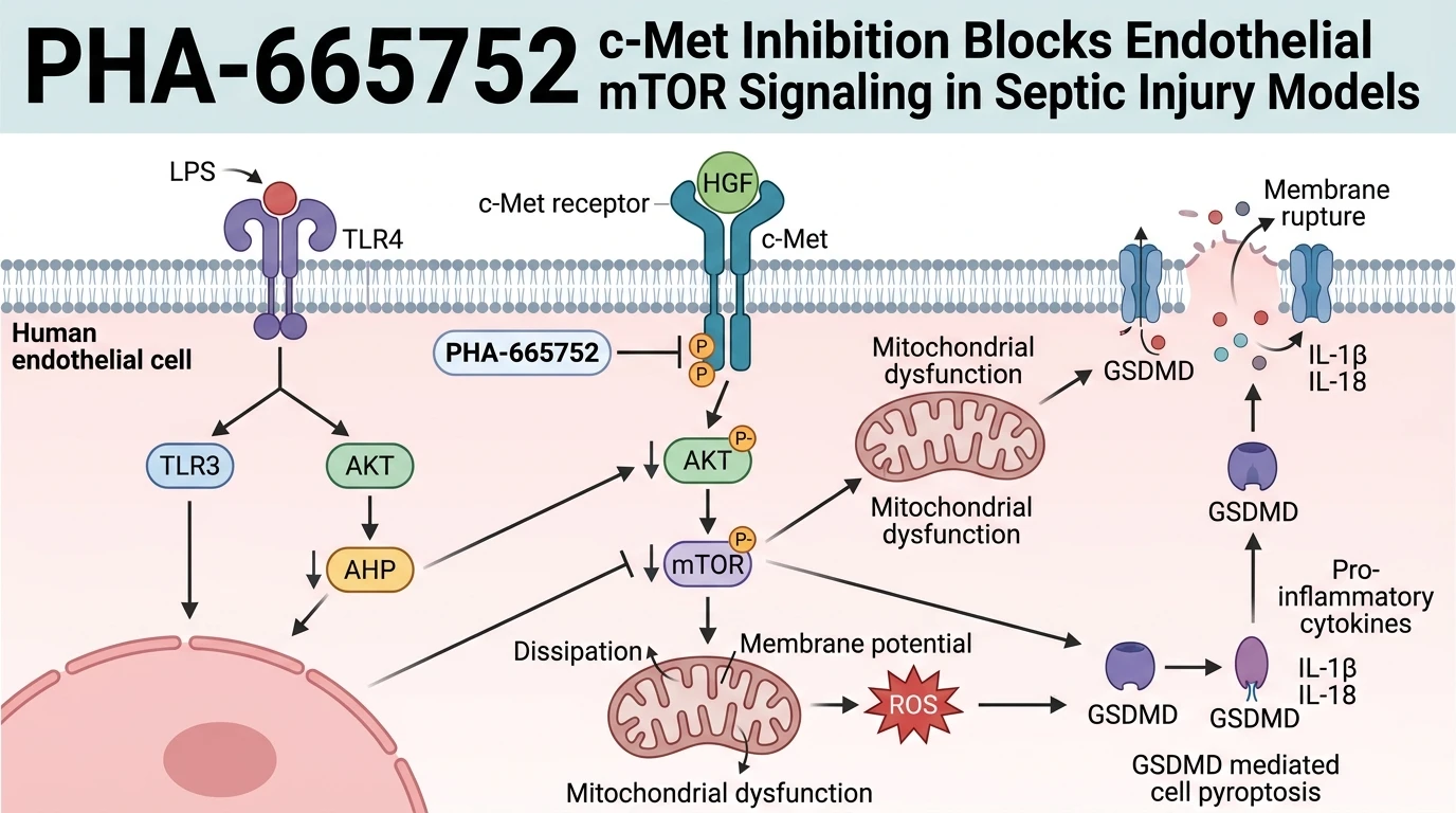
Endothelial cell dysfunction and microvascular disruption represent central pathological features of sepsis-induced acute lung injury. Lipopolysaccharide (LPS) exposure induces profound inflammatory damage in vascular beds. Hepatocyte growth factor (HGF) acts as an endogenous protective cytokine. Ligand engagement with its specific receptor, c-Met (Hepatocyte Growth Factor Receptor / Met Tyrosine Kinase), triggers receptor homodimerization and autophosphorylation across key catalytic domain tyrosine residues (Tyr1234/1235). This autophosphorylation creates high-affinity docking sites for intracellular adaptor proteins. Phosphatidylinositol 3-kinase (PI3K) recruitment subsequently activates downstream serine/threonine kinases. Through this PI3K-Akt signaling pathway cascade, AKT phosphorylates mammalian target of rapamycin (mTOR) at Ser2448, driving endothelial survival programs.
To dissect the mechanistic necessity of c-Met kinase activation during endotoxic stress, researchers employed PHA-665752, a cell-permeable, small-molecule ATP-competitive c-Met inhibitor. In cultured EA.hy926 endothelial cells transfected with LPS, exogenous HGF administration typically suppresses cellular injury. Receptor engagement by HGF limits gasdermin D-mediated pyroptotic pore formation, decreases intracellular reactive oxygen species (ROS) production, preserves mitochondrial membrane potential, and stabilizes monolayer barrier function. However, co-treatment with PHA-665752 abrogates these cytoprotective phenotypes. Enzymatic inhibition of the c-Met kinase domain prevents HGF-induced receptor phosphorylation. Consequently, downstream transmission along the c-Met/mTOR axis is completely suppressed under tested in vitro conditions.
Detailed biochemical assays demonstrate that PHA-665752 targets the ATP-binding pocket of c-Met with high nanomolar affinity. Inhibitor engagement prevents trans-phosphorylation of the activation loop. As a consequence, phosphorylation of downstream effectors, including AKT at Ser473 and mTOR at Ser2448, drops to undetectable levels in cellular lysates. Without active mTOR signal transduction, protective metabolic and anti-apoptotic programs fail to engage. Mitochondrial membrane potential depolarizes rapidly. Intracellular ROS levels accumulate, creating severe oxidative stress within the cytoplasm. Furthermore, execution of gasdermin D cleavage proceeds uninhibited, causing pore assembly in the plasma membrane and subsequent endothelial pyroptosis.
Vascular integrity metrics directly reflect these biochemical changes. In transendothelial permeability assays, treatment with PHA-665752 prevents HGF from maintaining tight junction integrity across EA.hy926 monolayers. Macromolecular permeability increases sharply following receptor kinase blockade. Electrical resistance across the endothelial barrier declines, mirroring microvascular leakage observed during severe septic shock. These experimental findings illustrate that c-Met kinase activity is essential for mediating HGF protective responses in septic vascular models.
Because these observations were collected within specific cellular assay systems, additional boundary conditions apply. In vitro endothelial monolayers lack physiological blood flow forces and organ-level cellular interactions. Additional cross-talk with heterologous receptor tyrosine kinases may occur in vivo. Nevertheless, pharmacological inhibition using PHA-665752 firmly establishes that c-Met catalytic activity and subsequent mTOR pathway activation are mechanistically required to protect vascular endothelial cells against endotoxin-induced pyroptosis and hyperpermeability.
Pharmacological Characterization of c-Met Blockade in Endothelial Models
Copy code to add this image to your page
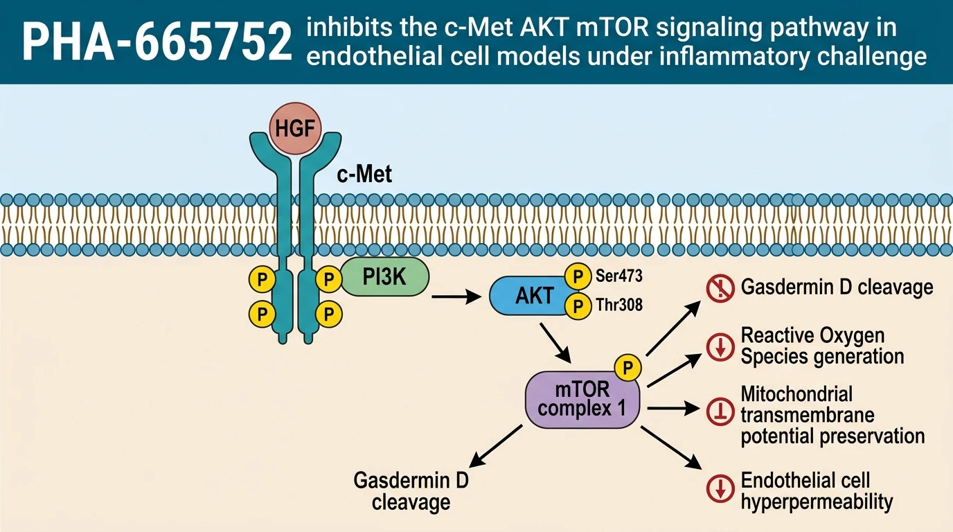
PHA-665752 acts as a selective, ATP-competitive inhibitor of the c-Met receptor tyrosine kinase. By abolishing ligand-induced c-Met autophosphorylation at Tyr1234/1235, the compound halts downstream activation of the AKT and mTOR signaling pathways in lipopolysaccharide-stimulated endothelial cells. Blocking this cytoprotective axis triggers mitochondrial dysfunction, exacerbates reactive oxygen species generation, accelerates gasdermin D-dependent pyroptosis, and increases vascular endothelial hyperpermeability. Thus, c-Met inhibition with PHA-665752 confirms that HGF mitigates septic endothelial injury via c-Met/mTOR signaling.
Abolition of c-Met and AKT Phosphorylation Cascades
Direct target binding was verified. In LPS-transfected EA.hy926 endothelial cell cultures, treatment with PHA-665752 completely blocked hepatocyte growth factor-induced autophosphorylation at c-Met tyrosine residues Tyr1234 and Tyr1235. Downstream phosphorylation ceased rapidly. Immunoassays showed that phosphorylation of AKT at Ser473 and mTOR at Ser2448 dropped significantly below baseline levels within thirty minutes of compound exposure. Protein expression levels remained stable. Pharmacological interruption at the receptor level directly decouples ligand binding from intracellular survival signal transmission under endotoxic challenge.
Exacerbation of Endothelial Pyroptosis and Permeability Metrics
Endothelial barrier integrity was compromised. Quantitative evaluation of cell death markers revealed that c-Met inhibition by PHA-665752 significantly enhanced lipopolysaccharide-induced endothelial cell pyroptosis despite exogenous HGF supplement. Gasdermin D cleavage escalated. Fluorometric assays demonstrated rapid dissipation of mitochondrial transmembrane potential, which directly triggered excessive intracellular reactive oxygen species accumulation within treated endothelial monolayers. Hyperpermeability was confirmed experimentally. Transendothelial electrical resistance values dropped, while macromolecular dextran permeability increased significantly in the presence of the small-molecule c-Met inhibitor.
Kinase Target Selectivity and Microvascular Boundary Conditions
Boundary conditions must be recognized. While these findings in EA.hy926 cell monolayers provide strong evidence for c-Met dependency, observations remain to be validated in complex animal models of acute lung injury. Cellular context influences signaling outputs. Under static in vitro culture conditions, secondary mechanical forces such as fluid shear stress and cell-matrix interactions are absent, potentially altering receptor transactivation pathways. Selectivity margins appeared consistent. Further investigation in randomized preclinical sepsis models will clarify whether systemic c-Met modulation impacts secondary pulmonary endothelial barrier function in vivo.